After an injury, medical providers often order diagnostic orthopedic imaging to see what has occurred inside of the body. Diagnostic imaging takes pictures of bones, ligaments, tendons, cartilage, muscles and more inside the body. These images help physicians determine how the injury has impacted the body to establish the best course of treatment.
The three main imaging methods are X-ray, magnetic resonance imaging (MRI) and computed tomography (CT) scan. Each method of orthopedic imaging offers a different view of the internal structures inside the body.
This article will compare the three main types of imaging for orthopedic injuries. Learn about the differences, advantages and disadvantages of an X-ray versus MRI versus CT scan.
X-ray imaging
What is an X-ray?
An X-ray, or radiograph, is an orthopedic imaging study that briefly sends safe amounts of electromagnetic radiation through your body to take images of the inside of the body.
X-rays are the most common kind of diagnostic imaging for orthopedic injuries. X-rays may not show as much detail as an MRI or CT scan, but occasionally they show the injury better than more sophisticated tests.
How does an X-ray work?
Following an orthopedic injury, the injured body part is positioned between the X-ray machine and the sensor. The patient holds still for a moment while the radiology technician takes the image. Patients often have multiple X-rays taken from different angles to provide the physician with the best picture of what has occurred in the body.
Occasionally, dye or another contrast material may be injected into a joint to outline soft tissues during an orthopedic X-ray. This is called an arthrogram. Arthrograms may also be used to determine accurate needle placement for fluid removal or injections into joints.
X-ray sessions typically last about 10 minutes. Images are often ready for the physician or radiologist to read quickly.
What do X-rays show best?
X-ray machines can quickly show images of orthopedic injuries like fractures, dislocations, misalignments or narrowed joint spaces. They also can help doctors diagnose infections, arthritis, osteoarthritis, bone cancer and more.
Orthopedic doctors use X-rays to monitor the healing process. They also use X-rays to evaluate the placement and alignment of implants or joint replacements.
Advantages of X-rays
- Wide availability
- Low cost
- Quickly performed studies – usually takes 10 minutes or less
- Quick turnaround of radiographic images
- Images help to diagnose orthopedic injuries like fractures and healing progress
Disadvantages of X-rays
- X-rays are less sophisticated than other types of orthopedic imaging
- Soft tissues are not as clearly visible as in other tests
- X-rays expose patients to low levels of radiation
- Special precautions must be taken for pregnant women to prevent harm to the baby
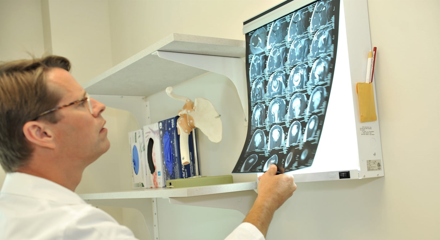
Magnetic resonance imaging (MRI)
What is an MRI?
Magnetic resonance imaging, or MRI, uses a powerful magnet to pass computer-generated radio waves through the body. MRIs create high-resolution images of the skeletal system, organs, soft tissues, nerves and blood vessels.
Instead of showing images on film like an X-ray, MRIs show cross-sectional images of the body. MRIs do not use radiation.
How does an MRI work?
During a typical MRI, the patient lies still on a table that slides into the tube-shaped MRI machine. The machine creates a magnetic field around the patient. It then sends computer-generated radio waves through the part of the body that is being imaged. The resonance of the body is recorded by a computer to create a highly detailed, two-dimensional image.
Cary Orthopaedics offers open-bore MRIs that allow patients to be less constrained and more comfortable during their procedures. This MRI experience is more like a CT scan and decreases feelings of claustrophobia.
MRI studies often take between 30 and 60 minutes to complete. The length of time for results to be received varies.
Since an MRI is a large magnet, patients with implants, joint replacements, pacemakers or other metal objects in the body must be evaluated to determine if the MRI can be performed safely.
What do MRIs show best?
MRIs provide detailed images of bones and soft tissues. They can be used to assess torn ligaments, tendons, cartilage and other soft tissue injuries. MRIs are often used to diagnose meniscus tears, rotator cuff tears, ACL injuries and spinal injuries.
MRIs also help physicians see the progression of arthritis and degenerative disk disease. Tumors, infections and herniated disks are other conditions often diagnosed with MRI imaging.
Advantages of MRIs
- Produce highly detailed images of soft tissues, bones, tendons and ligaments
- Can show subtle bone fractures, inflammation and tears of ligaments or tendons
- Differentiates between fat, water and muscle tissue
- No radiation is used
Disadvantages of MRIs
- MRI machines may be noisy, so patients have to wear ear protection or headphones
- The tube of the MRI machine may cause some patients to feel claustrophobic
- Patients must remain still for the full 30- to 60-minute test; movement during the test affects the image quality
- Not as common or readily available as X-ray machines
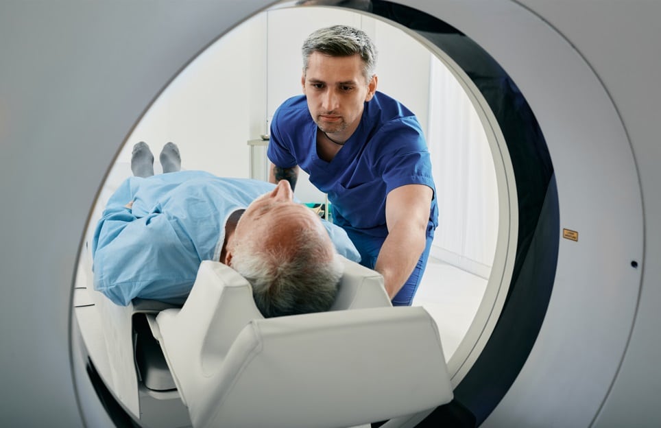
Computed tomography (CT) Scans
What is a CT scan?
A CT scan combines multiple X-ray images taken from different angles to create detailed, cross-sectional images of the body. CT scans provide 3-D images that help physicians diagnose and determine the size, shape and placement of structures in the body.
CT scans cost more and typically take more time to perform than regular X-rays. The time to receive the results of CT scans varies from an hour to more than a week, depending on how severe the injury is and other factors.
How does a CT scan work?
Similar to an MRI scan, the patient lies motionless on a table that slides into a CT scanner tube, but CT scanners are smaller than most MRI scanners. An X-ray tube rotates around the patient to take images from different angles. The images are combined on a computer to provide a 3D image. Some facilities offer open CT scanners.
Some patients may be given a dye or contrast to highlight a specific area during the CT scan.
What do CT scans show best?
CT scans offer a three-dimensional view of structures inside of the body, including bone and soft tissue. They show fine details of bone structures and can be used to diagnose muscle and bone disorders. A CT scan may be ordered by your orthopedic physician if other orthopedic imaging tests are inconclusive.
Advantages of CT scans
- Produce a 3-D view of injuries
- Detailed images of bones and soft tissues
- Useful to assess bones and joints when other orthopedic imaging tests are inconclusive
Disadvantages of CT scans
- More expensive than X-rays
- Takes more time than X-rays
- Less commonly used and available
- Uses radiation
Orthopedic imaging to diagnose your injury
When you or a loved one experiences an orthopedic injury, be sure to obtain the right diagnostic imaging tests. The results of the orthopedic imaging test will help the physician with accurate diagnosis and treatment of the injury.
The experienced orthopedic physicians and staff at Cary Orthopaedics have years of expertise in diagnosing and treating orthopedic injuries. We work to ensure that you receive the best orthopaedic imaging and treatment in the Raleigh area.


