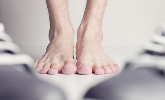
The human foot needs numerous functions to work effectively and efficiently. The anatomy of the foot can be divided into three main sections that include the forefoot, midfoot and hindfoot. There are joints, muscles, tendons and ligaments throughout these sections.
Cary Orthopaedics’ foot and ankle specialists focus not only on treating the symptoms of foot and ankle injuries but on understanding the underlying cause of the pain. We diagnose and treat a wide array of conditions, and we strive to educate our patients on the benefits of strengthening and stretching the feet and making proper shoe and insole selections.
A thorough understanding of every part is vital to diagnose conditions accurately. In this article, we highlight and explain the anatomy of the foot.
Bones
The foot is composed of 28 bones. In the hindfoot, midfoot and forefoot there are 11 types of bones that deserve a thorough explanation here.
Tibia and fibula
Two of those bones are the tibia and fibula that are responsible for connecting the foot to the rest of the body. The tibia is usually responsible for supporting up to 85 percent of the human body’s weight. The remaining 15 percent is distributed to the fibula. The primary function of the fibula is to be the lateral wall of the ankle mortise.
The tibia and fibula are held together by five different ligaments that are referred to as the tibiofibular syndesmosis.
Talus
The top bone of the foot is called the talus. It articulates with many of the other bones in the foot. Talus has three main parts: the body, the head and the neck. The body connects the talus to the lower leg.
The head is adjacent to the navicular bone to form the talonavicular joint. The talonavicular joint is seen as the universal joint of the foot. It allows for rotation, sideways movement, and up, down motion at the midfoot.
Then the neck serves as the entry point for many blood vessels supplying the talus.
Other bones in the foot include:
- Calcaneus
- Cuboid
- Navicular
- Cuneiforms (3)
- Metatarsals (5)
- Phalanges (14)
- Sesamoid Bones (2)
Ossicles of the foot
Some feet may contain a few additional or accessory ossicles/bones. These bones are seen as developmental variants, with around 40 variations having been discovered.
Joints
The subtalar joint
This joint functions as a sort of bridge between the ankle and the foot. It transfers loads from the foot to the tibia or vice versa.
The transverse tarsal joint
This joint isn’t seen as a “true joint.” However, it’s the combination of two different joints (the calcaneocuboid and talonavicular joints). It gives the midfoot the ability to move independently of the rearfoot.
There are over 33 other important joints that are contained within the foot.
Muscles
There are two muscle compartments that are located inside the lower leg. 1) The superficial posterior compartment. 2) The deep posterior compartment.
These muscles (including an additional two in the upper leg) are together known as the extrinsic muscles of the foot.
Superficial posterior compartment
The muscles within this compartment include the gastrocnemius, soleus and plantaris muscles. These form the triceps surae. The gastrocnemius is involved in plantar flexion of the ankle, while the knee is in extension. It also is involved in flexing the leg at the knee. The soleus is involved in plantar flexion of the ankle. The plantaris is a muscle that’s thought to be absent in around 10 percent of the population. It’s involved in plantar flexion of the ankle but plays a limited role compared to the other two superficial posterior muscles.
The deep posterior compartment
The muscles inside this compartment are the flexor hallucis longus, flexor digitorum longus, tibialis posterior and the popliteus muscles. The first flexor hallucis longus is involved with the flexing of big toes while having a limited contribution to plantar flexion of the ankle. The flexor digitorum longus aids the flexion of the four other toes on the foot. The tibialis posterior is mainly involved in ankle plantar flexion and also the inversion of the foot. Finally, the popilteus Muscles control weak flexion of the knee and also medial rotation of the tibia.
Ligaments and tendons
There are several ligaments in the human foot that support a lot of the human body and have important functions. However, this article will focus on just four of those ligaments.
ATFL
The anterior talofibular ligament is the ligament that is most commonly injured when ankles are sprained. The ATFL runs along from the lateral malleolus down to the front portion of the ankle connecting the neck of the talus. It’s responsible for stabilizing the ankle against inversion.
Posterior talofibular ligament
This ligament runs from the lower back of the fibula and to the outside back portion of the calcaneus. It functions to stabilize the ankle joint and subtalar joint.
CFL
The calcaneofibular ligament is also located on the lateral side of the ankle. It begins at the fibula and then goes along the lateral aspect of the ankle into the calcaneus. It resists inversion.
Deltoid ligament
The deltoid is actually a fan-shaped band of connective tissue on the medial side of the ankle. The ligament itself functions to support the resistance of eversion.
Some of the other ligaments that should be mentioned are:
- Spring ligament
- Lisfranc ligaments
- The inter-metatarsal ligaments
- The first joint capsule of the great toe (If this band gets stretched out, you could develop a bunion.)
- Anterior inferior tibiofibular ligament (Injuries having to do with this ligament are called high ankle sprains. They arise when the foot is fixed on the ground as the leg rotates inwards.)
- The interosseous membrane
- The syndesmosis
Tendons
There are over 100 tendons in the foot alone. They all work together to support your body, so it’s unnecessary to list them all individually. However, there is one that needs to be addressed in the anatomy of the foot.
The Achilles tendon
The Achilles tendon is the central tendon of the doot. It runs along with the calf muscle to the heel. This tendon is what makes it possible for you to run and jump, but also stand on your toes. It’s essentially responsible for lifting your heel off the ground when you undergo activity.
Foot and ankle care in Cary and Raleigh
Don’t underestimate the importance of having healthy feet. They are your body’s foundation, and vital to your overall health. If you or a loved one are having foot or ankle issues, or are interested in learning more about the anatomy of the foot, contact us today to meet with an orthopaedic foot specialist.


